Difference between revisions of "Surgical Procedure"
From NeuroTychoWiki
NaotakaFujii (talk | contribs) |
NaotakaFujii (talk | contribs) |
||
| Line 30: | Line 30: | ||
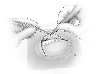
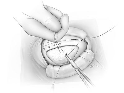
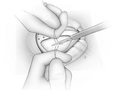
|[[File:Fig14.jpg|400px]] || Elevate dura with traction sutures. One suture is held by assistant and the other by surgeon. The traction sutures are used to enlarge subdural space to prevent damaging brain when opening dura. Touch dura with knife gently in between tractions and cut slowly layer by layer. Cutting doesn't have to be long. ~5mm will be fine. If you cut through dura, you will see transparent liquid (spinal fluid) comes out. But if you don't see the leakage, it is still on the way. | |[[File:Fig14.jpg|400px]] || Elevate dura with traction sutures. One suture is held by assistant and the other by surgeon. The traction sutures are used to enlarge subdural space to prevent damaging brain when opening dura. Touch dura with knife gently in between tractions and cut slowly layer by layer. Cutting doesn't have to be long. ~5mm will be fine. If you cut through dura, you will see transparent liquid (spinal fluid) comes out. But if you don't see the leakage, it is still on the way. | ||
|- | |- | ||
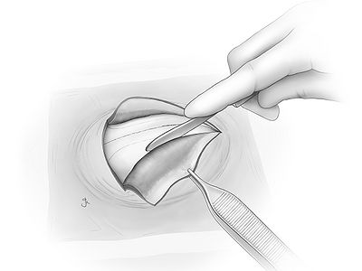
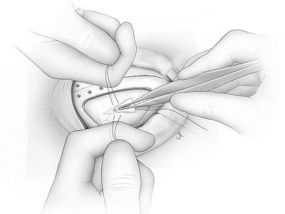
| − | |[[File:Fig15.jpg|400px]] || Cut Bensheet in triangle shape and soak in saline. | + | |[[File:Fig15.jpg|400px]] || Cut Bensheet in triangle shape and soak in saline. Insert the sheet into the dura hole gently. This will make safe working space for extending dura incision. |
|- | |- | ||
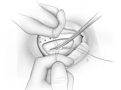
| − | |[[File:Fig16.jpg|400px]] || | + | |[[File:Fig16.jpg|400px]] || Cut dura with scissors. When cutting area moves, the sheet has to move together. Incision has to be always made above the sheet to protect brain. Don't cut too close to bone edge, it will make a difficulty when suturing. |
|- | |- | ||
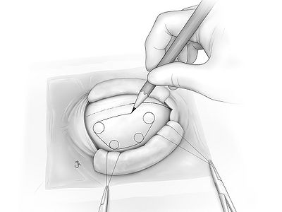
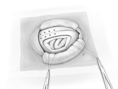
| − | |[[File:Fig17.jpg|400px]] || | + | |[[File:Fig17.jpg|400px]] || Brain is exposed. |
|- | |- | ||
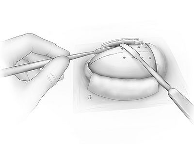
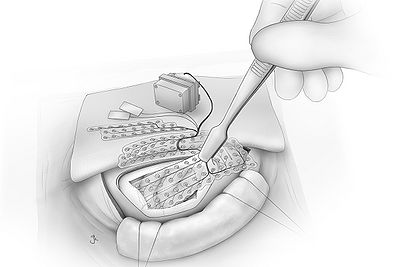
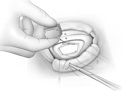
| − | |[[File:Fig18.jpg|400px]] || | + | |[[File:Fig18.jpg|400px]] || Insert ECoG array into subdural space. Use flat head forceps to hold the array. |
|- | |- | ||
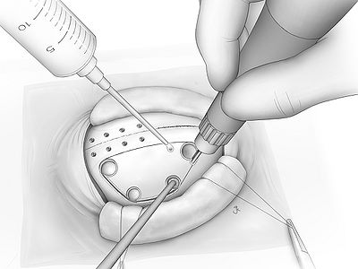
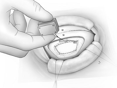
| − | |[[File:Fig19.jpg|400px]] || | + | |[[File:Fig19.jpg|400px]] || Place reference electrode in subdural space (between ECoG sheet and dura) and ground electrode in epidural space (between dura and skull). |
|- | |- | ||
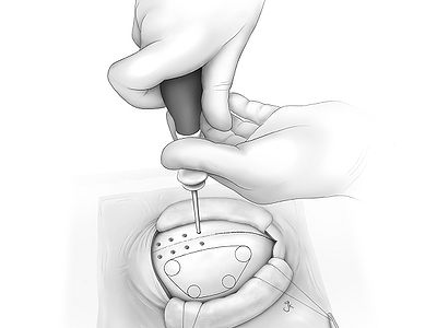
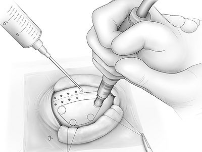
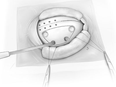
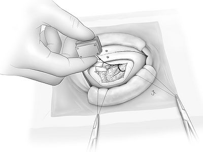
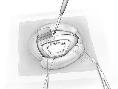
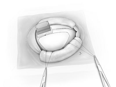
| − | |[[File:Fig20.jpg|400px]] || | + | |[[File:Fig20.jpg|400px]] || Cut artificial dura that fits to the size of dura opening. Insert artificial dura in subdural space. Rim of the artificial dura has to be covered by dura. Put sutures(PBS: a thread made of biodegradable plastic) at corners. |
|- | |- | ||
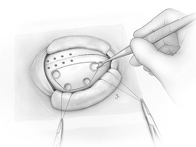
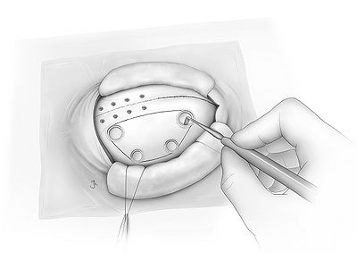
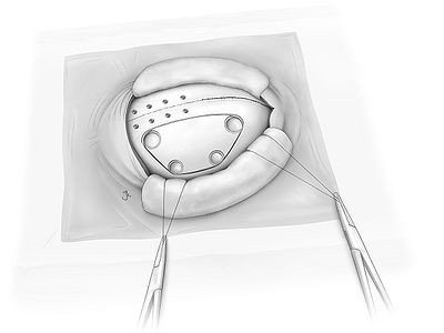
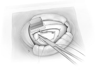
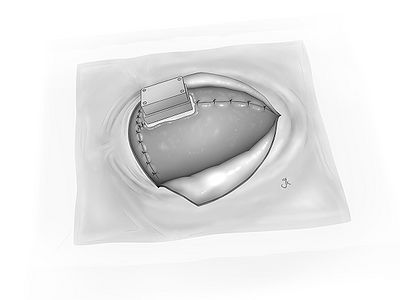
| − | |[[File:Fig21.jpg|400px]] || | + | |[[File:Fig21.jpg|400px]] || Each rim is sutured by uninterrupted suture. |
|- | |- | ||
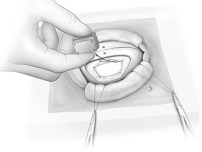
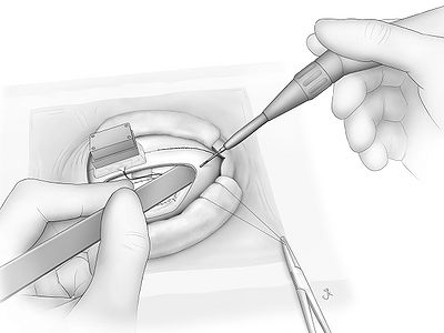
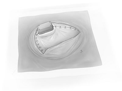
| − | |[[File:Fig22.jpg|400px]] || | + | |[[File:Fig22.jpg|400px]] || Wrap a hole where wires are coming out from subdural space with small piece of fascia and suture it to dura securely for preventing spinal fluid leakage. |
|- | |- | ||
|[[File:Fig23.jpg|400px]] || (figure 23) | |[[File:Fig23.jpg|400px]] || (figure 23) | ||
Revision as of 09:27, 14 August 2011
Surgical illustrations courtesy of [Juna Kurihara]

